Some say the process of underdevelopment, begins long before conception since it depends on the
primary cells and their conditions so that it can develop properly the Nervous
System Central
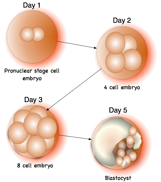
The race begins with the meeting
between the sperm and the ovule. Both cells must contain a specific genetic
load and determined to prevent hazards effacement, you overlap, mosaicism or
lack of alleles or genetic material elsewhere. Both cells combine their
material, resulting in a single cell.
Also it is known that there
must be a correct proteinaceous load to ensure the success of the process design.
So that what generates the
diversity of races and physical features is genetic recombination which
undergoes each generation, but each individual is genetically different from
everyone else (except if you have an identical twin), since the variety of eggs
or sperm that are formed along the life is so great that for practical purposes
only can say that none of them is equal to the other. Thus, mutations are the
raw material of genetic diversity, but is even greater and less controllable in species
with sexual reproduction, facing all the time different genomes .
Subsequent to this process is
said that the Meiosis which is a process of cell division in which a diploid
cell (2n) undergoes two successive divisions, with the capacity to produce four
haploid cells (n).
This process is carried out in two divisions
nuclear and cytoplasmic, called first and second division meiotic or simply meiosis
I and meiosis II.Both are part of of the prophase, metaphase, anaphase and
telophase.
In the interface is
duplicated genetic material is shared while that homologous chromosomes are
divided into two daughter cells in meiosis I , the phenomenon of
cross-breeding.
Once you pass this stage, it
is possible the beginning of meiosis II, like in a mitosis, each
chromatid migrates to a pole. The result is 4-cell daughters haploid (n).
During meiosis are matched
member of each homologous pair of chromosomes during prophase, forming
bivalent. During this phase it developed a protein structure called the
synaptonemal complex, allowing recombination between two homologous chromosomes
that occurs during this phase.

Subsequently a large
chromosomal condensation occurs and the bivalent are situated on the equatorial
plate during the first metaphase, resulting in the migration of n
chromosomes to each of the Poles during the first anaphase.
This reduction division is responsible
for the maintenance of the characteristic of each species chromosome number.
In meiosis II, the chromatids
that form each chromosome separate and are distributed to the daughter cells
nuclei. Between these two successive stages there is no stage S (DNA
replication). The maturation of the daughter cells gives rise to the gametes.
Something important to note in
this regard is that the genome of a human normal consists of 23 pairs of
chromosomes, the inherited by mother and father inherited that form each pair,
but in total there are 24 pairs of chromosomes that 2 correspond to the sex
chromosomes X and the and, which combine in XX if you are female and XY if it’s
a male.
All this takes place in a
relatively short period of time and in spite of being a process necessary for
the reproduction of the human species, is not a perfect process; errors in
meiosis are sometimes responsible for the main chromosomal anomalies. Meiosis
manages to keep constant the number of chromosomes in the cells of the species
to maintain the genetic information. In general, members of a chromosome pair
are not in close proximity either at rest or during mitotic division cell. The
only time they enter into intimate contact is during the meiotic divisions or
germ cell maturation.

This process continued during
the following weeks the cells begin to migrate and give way to another process
called referred to as phase of cell proliferation to one in which the cells
that compose the Nervous System (neurons and glial cells) originate or are
born.
Of the different stages of
Morphogenesis is this which can properly be considered as the phase of
neurogenesis.
Since it is known that the
development of the human brain starts very early, around 3 to 4th week of
gestational age and continues, although at a declining rate, until adulthood.
And this development is characterized by the occurrence of 2 major
organizational events.
The first begins with the
conception and includes neuroregulation events, proliferation, migration, and
differentiation, the second occurs after birth. It has been proposed that these
events are controlled by genetic factors and epigenetic (non-mutational phenomena
but that vary the expression of a gene, such as the development of proteins or
blocking of certain neurotransmitters) that originate neural structures
sensitive to external influences.

In humans this stage of
development occurs in the fourth week of gestation from the neuroepithelium, which is made up of the calls of
CNS stem cells. This stem cell progenitor, which also glioblasts or
immature neurons produce called cells. Once born neurons, that as it has been
said are still immature, they lose their reproductive ability. The glioblasts,
however, retain their reproductive capacity throughout life.
This phase covers until about
the fifth month of gestation; although we cannot forget that it does not occur
simultaneously in all neural tube, but that each region has its own period of
neurogenesis. The process does not end there, but rather so that we can
properly talk of nervous system cells that compose it still must go through
different times.
After this phase of cell
proliferation occurs cell migration, in which nerve cells migrate to their
final location; the radial glia is the support through which neurons can reach
their final location.
Cells in these phases are
still undifferentiated, so go to the stage of neuronal differentiation to
acquire the morphological and physiological characteristics of the mature
neuron. Also, establish different connections (synapses), while the development
establishes many more synapses than necessary during synaptogenesis, with which
many of these connections are subsequently eliminated. In addition, during
fetal development the human creates many more neurons than needs, so those that
are functionally superfluous die (this neuronal death is known as neuronal
apoptosis and can reach between 25% and 75% of neurons created).

It is so nervous tissue
formation begins with the formation of a simple tube, the so-called neural
tube and from the induction of the neuroectoderm (this is part of the
ectoderm that is the outermost cell primary embryo
that originates the central and peripheral, nervous systems including some
glial cells), this process occurs in the human between the third and fourth
gestational week.
Once formed the neural tube
occurs a differentiation in three dimensions: the first leads to the spinal
cord, the second will give rise to stem and brain stem and the cerebellum,
while the third portion will develop the cerebral hemispheres. This stage is
called a fore brain, this process that
occurs between the fifth and tenth gestational week and during which develops
an active neurogenesis (neuron development) from neural precursor cells, which
have a special feature and is not mature and do not proliferate, because we
will have to wait for the next moment for such differentiation.
 Between the eighth and eighteenth gestational week, occurs an active
neuronal proliferation, the precursor cells begin to differentiate to produce
new precursor cells and neuronal cells such are different neurons as glial
(cells astrocytes and oligodendrocytes).
Between the eighth and eighteenth gestational week, occurs an active
neuronal proliferation, the precursor cells begin to differentiate to produce
new precursor cells and neuronal cells such are different neurons as glial
(cells astrocytes and oligodendrocytes).
Neuronal migration occurs mainly in two regions in the thalamus and
hypothalamus, where the oldest neurons are pushed by more new neurons, by which
the first will be located in the periphery.On the other hand, in regions of the brain structure of laminar, as it
is the case of the cortex and the cerebellum, neurons more young people migrate
to break through to the oldest, whereupon the latter will sit closer of the neuroepithelium
and the more young people on the periphery.
Neuronal migration process
takes place between the 10th second and the twenty fourth gestational week.
During neurogenesis and
neuronal migration, approximately 50% of neurons undergo apoptosis, i.e.
die in a programmed way, probably because they do not follow the correct course
of emigration or because they do not receive adequate stimuli, the correct
answer is still a mystery.
A certain proportion of the
neurons that survive (20%) Trek horizontally and one after emigration radial,
to allow the formation of lamination (segmentation) cortex, it is so neurons
looking his way, motivated by chemical stimuli (Neurotropic factors), extending
its structure in one of its ends, resulting in the so-called axonal growth cones
.
Simultaneously with the
neuronal migration occurs in synaptogenesis (formation of synapses),
although this is much more intense between the twelfth and the twelfth fourth
gestational week, but persists in a very active way until the eighth or ninth
month post natal.
It is interesting to note that
pre natal synaptogenesis is mainly determined by the genetic heritage of the
individual. However, in the stage post natal synaptogenesis is also affected by
sensory experiences, particularly through the learning process.
Thus, during puberty, occurs a
sort of freeze on neurogenesis, which has been associated with the acquisition
of the own and particular character of each individual. Myelination is a late
process that starts in way more intense from the 40ava week, occurs in the
white matter and peripheral neurons
Neurogenesis and the
subsequent stages associated with this process morphogenic lead to the
formation of approximately 100 billion neurons in the adult brain and several
trillions of synapses.
This implies that a
significant number of the 30,000 genes that we have must be involved in this
complex process, expressing together in simultaneous or sequential form.
However, she has still not been achieved understand this prodigious process,
because a region possessing 20,000 genes, is only 302 neurons and nerve tissue
that form is far from having the functionality of the human brain.
The number of cells in the
fetal brain is between 30 and 70% higher than the number of neurons in the
adult. Surplus cells survive for a period of days to weeks, after which, on its
own, starts a cascade of degenerative changes and a physiological process of programmed
cell death.
In the picture below, it is
possible to observe the differences between birth and two years of development,
although it seems that increased neuronal tangle, in reality there are what are
they are less neurons with larger number of neural networks, connections
between neurons, i.e. interneuronal communication, which allows a more robust
network that ensure more specific skills.

In this sense, found that the
selective removal of the synaptic connections, is a fundamental process in the
cognitive development of the child, as has been observed relationship between
changes in the gray matter of the frontal lobe and the evolution in the
performance of cognitive tasks.
During the acceleration phase,
occurs a large increase of dendritic extensions and small branch, which has
been called dendritic arborization, that form numerous synapses, so that
all cells and its extensions are arranged in layers and orient themselves, at
the same time causing programmed cell death and differentiation and
specialization neuronal This depending on the interactions with the environment
and genetic factors. So crests of the neuronal branches are, density peaks
occur at different ages, but also in different brain areas.
Thus one fast and dense
development both in the visual cortex and the hearing between the 3 and 4
postnatal months and maximum density, around the year of life can be observed.
On the contrary, the growth of the prefrontal area is presented at the same
age, but the peak is reached until after the first year of life. The only
exceptions are granulated cells of the olfactory bulb, cerebellum, and
hippocampus, which continue its genesis after birth and continue throughout
life.

Brainly, myelination,
that is an overlay of the neural connections by a
specialized membrane which allows a proper transmission of nerve impulses, is
fundamentally a made post natal, occurring in cycles, with a ranked stream by
default, to thus start the neural connections, the most important, which
will form the basis for all subsequent development.
Thus, myelination greatly
contributes to improve the functionality of the brain because it produces an
increase in the speed of nerve impulse conduction. In this sense has been found
that there is an increase in white matter during childhood, which probably
reflects the increase in myelination.
References:
Álvarez Buylla, A. & García Verdugo, J.M. (2002) Neurogenesis in adult subventricular zone. Journal of neuroscience. 22 (3) 629 - 634.
Avaria, M. A. (2005) Aspectos biológicos del desarrollo psicomotor. Rev. Ped. Elec. [en línea] Vol 2, N° 1.
Bloom, F: Beal, M & Kupfer, D. (2006) The Dana guide to brain health. Dana Press. Estados Unidos.
Coplan J. (1985) Evaluation of the child with delayed speech or language. Pediatr Ann. 14: 203-8.
Deacon, T. (2000) Evolutionary perspectivas on language and brain plasticity. Cognitive science. 28 (1) 34- 39.
Flores, J. (2005) Atención temprana en el síndrome de Down: Bases neurobiológicas Rev Síndrome de Down 20: 132-142.
Gage, F. (2007) Brain, repairs yourself. In Floyd E, Bloom (2007) The best of the brain from Scientific American: mind, matter, and tomorrow’s brain. Washington DC. Dana Press.
Gollin. E. S. (1981) Developmental and plasticity: behavioral and biological aspects of variation in developmental. New York. Academic Press.
Kaplan, B. A. (1983) Developmental psychology: historical and philosophical learning. New Jersey. Elrbaum Hillsdale.
León Carrión, J. (2003) Células madre, genética y neuropsicología. Revista Española de Neuropsicología. 5 (1) 1-13.
Maciques (2004) Plasticidad Neuronal. Revista de neurología. 2 (3) 13-17.
Morgado, I. (2005) Psicobiología del aprendizaje y la memoria. Cuadernos de Información y Comunicación. 10. 221- 233.
Nieto, M. (2003) Plasticidad neural. Mente y cerebro. O3. 72-80.
Poch, M.L. (2001) Neurobiología del desarrollo temprano, Contextos Educativos. 4 79-94.
Thanks for this excellent post!
ReplyDeleteThank you so much for taking a moment to write a ocmment!. I wrote this for a course about neurodevelopment and brain plasticity but I feel happy it can be good for the public!.
DeleteThanks for reading it!!
Very detailed, well structured and bursting with information.
ReplyDeleteKeep this up,
Brendan.
Thank you so much for your comment Brendan!!
DeleteI really appreciate it!